
Year 12 Biology Students Study Mitosis in Garlic Root Tip
Year 12 Biology class has been actively investigating the process of mitosis in garlic root tip cells.
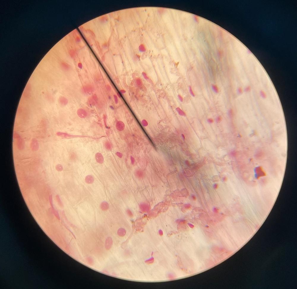
Garlic bulbs were sprouted, and root tips were collected, stained with acetic orcein, and then observed under a microscope at 400 times magnification. The photograph above depicts the garlic root tip cells, where the large pink blobs represent the cell nucleus, and the faint pink outlines indicate the observable cell walls.
Upon closer inspection, centrosomes and early miotic spindles were visible, suggesting the occurrence of the Interphase and Prophase stages. Unfortunately, no cells in Anaphase were observed that are characterised by distinguishable spindle fibres.
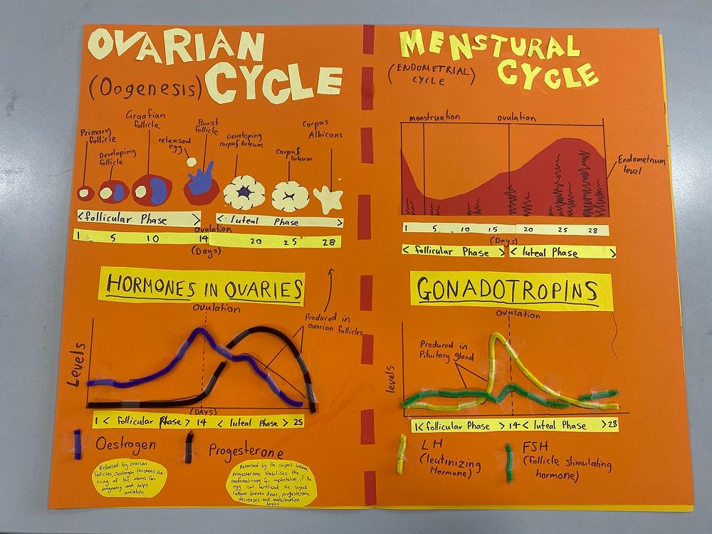
The students have also been learning about the menstrual cycle and ovarian cycle. Through a combination of theoretical study and hands-on activities, students have gained a comprehensive understanding of the hormonal dynamics and physiological changes that characterise these cycles. They have explored key phases such as follicular development, ovulation, and the formation of the corpus luteum in the ovarian cycle, as well as the corresponding changes in the uterine lining during the menstrual cycle. Above is a poster that was created by the talented Year 12 Biology student Emily M-A.